Saturday, 13 November 2021

Where is Your Focus? Let's Play Spot the Ball.

I think that the hunt for a focus of infection in a child is a lot like a game called “Spot the Ball” in which people looked at a picture from a football game with the ball removed and tried to guess where the ball was.

Finding a focus of infection is a very interesting topic at the moment.  More than ever, primary care clinical assessments are occurring remotely rather than face to face. Febrile children are being assessed without laryngoscopy or auscultation.  This seemingly contradicts the tradition of the need to find a focus of infection.  So what is the deal?  I've been asked a lot of questions about this recently and I thought that the simplest thing to do was to bring together my answers.

Do I need to find a focus of infection?

No.  There is no absolute need to find a focus of infection.  Here are a few facts that disprove the idea that a febrile child needs a physical clinical assessment for focus of infection in every case:

  • If finding a focus was mandatory, parents and carers would have to have their febrile child assessed on every occasion.  Self-care without medical assessment would be neglectful and therefore a safeguarding issue.
  • Even if you believe that medical assessment should take place, this only provides a snapshot.  Childhood febrile illnesses are dynamic and the focus can change.  A child who has an otitis media now could have mastoiditis before the day is over.  If we always need to know the focus, you would never be able to send the child out of your sight.

Does finding a benign focus rule out serious infection?

No.  That’s not how that works.  To illustrate the point, I'll tell you a story.  A 4 month old child came in with a history of fever and irritability progressing to vomiting and unresponsiveness.  On assessment they had a GCS of 8, heart rate of 200 and capillary refill time of 4 seconds.  I looked in their ear, found that they had otitis media.  We all celebrated the finding of the focus and discharged the patient with oral medication. 

That would never happen of course.  While I give that as an ridiculous example, the principle does apply to the moderately unwell child also.  Finding a focus can be a distraction or a premature conclusion.  If we are misled into thinking that an upper respiratory focus is the end of the decision making, we're missing a bigger picture.  We still need to step back and look at the overall clinical scenario.  So in the need to rule out things like meningitis, sepsis and pneumonia, finding a focus could be diagnostic noise.  Serious infections are ruled out on their own merits, not by finding another focus.

Does a runny nose count as a focus of infection?

Yes, but the question of infection doesn't work like that.  A child with a runny nose and a non-specific cough can be presumed to have an upper respiratory tract infection. The real question is, do they have features of another more significant focus?  If they have difficulty breathing, are seriously unwell or specific features of something else (e.g. reduced conscious level) then the assumption flicks from "presumed uncomplicated URTI" to "presumed complication of URTI".

One of the values of giving credibility to non-specific respiratory symptoms is that it helps to answer the age-old question of "should I get a urine sample from this child?"  That's a whole new can of worms.  In a massively oversimplified answer, if they are well and have a cough and runny nose you don't need one.  If they have no cough or runny nose you do need one, especially if they have abdominal pain, vomiting without diarrhoea or have strong smelling urine.

So if focus can be misleading and can change soon after I've assessed it anyway, what am I actually supposed to be doing? 

Treat the assessment of an unwell child like a game of spot the ball.  There will be clues and distractions.  There will be things that direct and misdirect.  The main task is to look for signs of a focus that needs to be treated.  Uncomplicated upper respiratory tract infection (including tonsillitis and otitis media) are not on the list of things that must be identified and treated.  What is interesting is that uncomplicated lower respiratory tract infection’s (LRTI/ pneumonia) place on the list is increasingly in question.

What is on the list of significant infections and how do I detect these?

  • Complications of URTI are rare but significant.  Externally, mastoiditis and lymph node abscess can be detected clinically.  Internally, peritonsillar abscess should be looked for in a child who is unable to swallow despite adequate analgesia.
  • Central nervous system (CNS) infection (meningitis, encephalitis, abscess) disproportionately affect function and behaviour.  Reduced or focally abnormal CNS function is a red flag.  Signs and symptoms are often non-specific in children but the non-specific signs such as inability to settle are persistent in a way that is unusual for uncomplicated infections.
  • Pneumonia, when accompanied by significant respiratory distress or systemic effects is still going to be on the list.  The fact that there is mounting evidence that pneumonia without significant effects does not mandate treatment simplifies the assessment of focus.  Localised signs on auscultation is a focus but the need for antibiotics in the absence of more significant findings is questionable.
  • Urinary tract infection is a paradoxical focus in children.  While UTI can self resolve without significant likelihood of complications, these low grade infections often go undetected.  The UTIs that are clinically apparent in children are the most severe UTIs.  It is therefore normal to treat all UTIs especially in pre-school children.  Urine samples should be tested if there is no clear focus elsewhere.  In the child under three years old, ideally these samples are sent for microscopy and culture.
  • Finally, there is the focus that is not actually infection.  Inflammatory diseases such as Kawasaki and PIMS-TS present with 5 or more days of unrelenting fever, mucousitis and a very miserable child.  Consider this possibility when the child is worse or no better on day 5 of a febrile illness.

When you play spot the ball in the traditional way, you don't get to find out if you were correct until the next day.  What could be more like hunting for the serious focus amongst the hundreds of febrile children you will see or assess remotely this year?

Good hunting.

Edward Snelson
@sailordoctor


Thursday, 2 September 2021

With every crisis an opportunity - How to use blood count in children

Another week, another crisis - This time, in the UK we have a major shortage of blood bottles.

Primary care have been advised to suspend non-urgent blood tests for the next few weeks during a major shortage in sample bottles. Personally, I blame the 5G mast they put up near the hospital.

When we are faced with limited resources, we have to get even better than ususal at decision making.  We have an opportunity to look with fresh eyes and recognise those areas of practice which may have become facile and revisit how we use available resources.  In this post, I'll be looking at the use of blood counts in children.  When are these tests likely to be genuinely helpful and when are they unlikely to be the best next step?

First of all some general principles:

The last one in that list is particularly relevant to the question of when to do a blood count.  

A blood count gives a wealth of information about various haematological parameters.  Because there are so many being reported, it is reasonably likely that any report will include one parameter that is outside of the statistical norm.  One of the reasons for this is that children have very active and responsive bone marrow and immune systems.  Peaks and troughs in white cells are more common and extreme in children even if well.  As a result, blood counts can be historical by the time they are reported.


It is very common to find an abnormal parameter (e.g. lymphopaenia) for which the recommendation is simply “repeat”.  By the time the result is known by the requesting clinician, it may well be the case that the child has made plenty more of the lymphocytes in response to the dip reported by the test.


Of course, chronic and persistent abnormalities do exist.  The next question is, are these usually a surprise finding on a blood test?


The simple answer is no.

  • Congenital immunodeficiency is extremely rare in children and does not usually present as a chance finding on a blood test done to investigate low-level concerns.  It usually presents with severe and atypical infections and does so early in life.  
  • Acquired haematological problems (HIV/AIDS aside) will usually present with significant systemic signs and symptoms – chronic fatigue, pallor, weight loss or pyrexia of unknown origin.

It is notable that it is exceedingly rare for children with cancer to present via the two week wait referral pathway(1) that exists in the UK.  Instead, the vast majority present via the ED or as an acute referral from a clinician in Primary Care.  The likely reason for this is that there are red flags apparent to the parent or the primary care clinician.  The two week wait pathway tends to be used in cases where the child has a worrying feature without serious red flags.  (e.g. a solitary palpable lymph node which is not growing and in the context of a well child)  In other words, when a child is pale and lethargic and looks unwell, someone makes sure they are seen immediately.  Serious illness usually presents with atypical signs or symptoms.


This brings us onto another rule of thumb:

The decision to perform blood tests is sometimes a sign of uncertainty.  The clinician feels unable to be completely reassured yet is not able to reach a definitive diagnosis.


To illustrate this, let me give you a clear haematological example of a blood test being done to answer a specific question: 

A child presents one week after a viral illness.  They are well and afebrile but now have a diffuse petechial rash.  There is no lymphadenopathy, pallor or hepatosplenomegaly.  The clinician considers the possibility that the child has immune thrombocypaenia purpura and does a blood count to check platelets.  The blood test confirms the diagnosis.


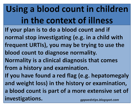
If a blood test is done without a specific question, or because the clinician hopes that the blood test with add weight to a diagnosis of normality, this is problematic.  Due to the above mentioned fluctuations, there is a high probability that the result will not be completely normal.  If the aim is to gain time, watchful waiting without a blood test is often the more valid approach.

The current crisis may be the perfect opportunity to ask questions about the role and importance of blood tests in children.  When it comes to blood counts in children, these rarely give useful information in the absence of a pre-test clinical sign or symptom which already gives a very high probability of a haematological or immunological problem.  Using a blood count to check something specific such as haemoglobin is a far more precise science.   Ask a question, get an answer.  Boom.

Edward Snelson

@sailordoctor


Disclaimer - Human factors may influence the decision to do a blood count.  I have not included this in my analysis.  Draw your own conclusions as to why this may be, human.

References

  1. Roskin J, Diviney J, Nanduri VPresentation of childhood cancers to a paediatric shared care unitArchives of Disease in Childhood 2015;100:1131-1135.

Monday, 16 August 2021

Trial by Inhaler - Bronchiolitis vs Viral Wheeze

With wheeze in children becoming a major presentation again, it feels like a good time to explore the issue of deciding whether a child has bronchiolitis or viral induced wheeze.  There are various way that people do this in practice.  Many stick to a strict 12 month cut off.  This method works reasonably well and is rarely problematic.  Bronchospasm is rare below this age and if it is going to be problematic under the age of 12 months, in my experience the infant is severely distressed and gets bronchodilators out of desperation rather than a diagnostic trial.


I have already explored a method of determining whether the pathology causing wheeze is predominantly wetness (bronchiolitis) or tightness (viral induced wheeze/ bronchospasm) by using age combined with the story.

Slow accumulation of moisture and mucous tends to cause worsening of symptoms over days whereas bronchospasm causes acute change over hours.  My opinion is that in the majority of cases, the age and the story will correlate.

Where the patient is in the overlap zone (e.g. 10-15 months old) and the story is clear (e.g. snotty/ coughing on Monday, struggling with feeds on Tuesday, noisy breathing on Wednesday and fast breathing on Thursday) then the story gives the diagnosis.  With age/ story correlation or where the age allows ambiguity but the story is clear, the diagnosis is made.

So what about simply trying an inhaler to see if it works?  This alternative approach to the age of overlap sounds straightforward and is reasonably common in practice, but is it logical?

A therapeutic trial works best when a clinical effect is guaranteed and unambiguous.  Neither of these things is true in this situation.  With viral wheeze, which should respond to salbutamol, clinically apparent response may require increased or repeated doses.  Bronchiolitis, which will not respond, is famous for mini-fluctuations in work of breathing.  This is caused by mucous plugging or the clearing of secretions.

When you think of it in these terms, trying an inhaler doesn't meet the quality standards required of a valid test.

Trial by inhaler is also problematic due to human bias.  Uncertainty is fertile ground for biases to mislead us when an inhaler is given to make a diagnosis rather than as treatment.  It is better to use beta-agonists therapeutically where appropriate and to see non-response as a reason to reconsider a presumed diagnosis of viral wheeze.  If viral wheeze is the problem, we should not allow the lack of effect to refute the diagnosis.
Edward Snelson
@sailordoctor

Disclaimer - when I wrote this, I briefly thought that you could bring logic to medicine.  I know, right?!?

Friday, 21 May 2021

Joining The Dots - How to recognise the seriously unwell child

In the previous post, I outlined how to tell the difference between abnormal signs that are part of a functional and fully compensated response and those that are part of an illness that is having a more significant clinical effect.

The child who is completely well and the child who is visibly seriously unwell and decompensating are both fairly straightforward scenarios.  The child who is very well causes no clinical concern.  The child who is decompensating causes unambiguous and immediate clinical concern.  If only decision making was always this easy.

The child who is febrile with tachycardia but a reassuring level of activity and interaction is also relatively straightforward.  The trouble is that we end up having to make decisions about those children who have a reduced activity level or whose interaction is not completely reassuring.  Often there is a disconnect between what guidelines tell us to worry about and how worried we actually are.  I believe that this often occurs when we intuitively include something in our decision making that is rarely featured in guidelines: the pattern of the illness.

Everything written tends to be geared towards the snapshot:

  • What is the heart rate?
  • What is the temperature?
  • How active and interactive is the child?

This is problematic in any acute specialty assessing febrile children.  Catch the child at the wrong time and they seem to trigger multiple red flags.  Base your assessment on the snapshot alone and you may be falsely reassured.

As covered in the previous post, physiology in young children (not so much babies and older children) responds to illness with what can be dramatic changes even in uncomplicated low-risk infections.  Unfortunately the same abnormalities can be seen in more clinically significant infections.  No one thing is particularly sensitive or specific when deciding whether to be worried.

So if a snapshot can be misleading and there is significant overlap between low-risk and high risk scenarios, how can we decide when a clinical presentation is high risk for sepsis or serious bacterial infection?

The pattern of symptom progression is probably the answer.  In the past, research has concentrated on the snapshot, over-emphasising the assessment of various parameters at a single point in time.  Human intelligence allows us to incorporate the more complex business of considering three very important factors that previous research has not often considered or emphasised:

  • The most recent worst state of the child
  • The most recent best state of the child
  • The pattern and progression of symptoms over time

The evaluation of the best and worst states and the pattern may be include retrospective (history) and prospective (a period of observation) information.  The likelihood is that you already put a great deal of weight on these factors in your decision making.  It is also likely that you have recognised that there are two main patterns of illness in children.  I have represented these in the following diagram, without labelling which is the low risk scenario and which is high risk.

Intuitively, I would assume that you recognise the dotted line as the high risk pattern and the solid line as the typical pattern of a low risk clinical picture of childhood febrile illness.

I suspect that the explanation for this is that the child represented by the solid line is exibiting a physiological reponse to an infection that does not cause organ dysfunction.  Therefore as their immunological response fluctuates, they swing from one extreme to the other.  The child represented by the dotted line is sufferng from an infection which is causing significant physiological dysfunction.  The result is an inability to return to normal.

If you buy that, have another look at the diagram and look at the lines in the first half from a time point of view.  In the left hand part of the diagram, severity of symptoms is often worse for the child who swings from being more unwell to a return to baseline.  I believe that this is the reason that formulaic risk-assessment for serious illness that is based on a snapshot is impractical and problematic.

The end results of this over-emphasis on clinical information taken at a single point in time are twofold:
  • Using a snapshot will over-diagnose serious illness, simply due to the pre-test probability of sepsis and serious bacterial infection (SBI) in the low-risk (which is most children) child.
  • Over time, the repeated realisation that children usually have an uncomplicated, self-limiting illness despite the severity of symptoms and abnormal physiological parameters risks de-sensitises the clinician to the possibility of sepsis/SBI.  Abnormality becomes normalised and we learn to ignore things that have poor specificity for a serious outcome.
I would strongly encourge stepping back from the snapshot to see the big picture over time.  Doing so, either through the history, observation or both might help you to recognise which children are best managed symptomatically and with good safety netting advice.  It migh also allow earlier recognition of the less common scenario of the child who persistently fails to return to (or close to) baseline and is more likely to have a more significant illness.

Telling the difference between self-liniting childhood illness and serious infection is complex and requires us to process an awful lot of information.  When it comes to features like best and worst states and the pattern of symptoms over time, your intuition is probably already joining these dots for you.  I think you should trust that intuition.

Edward Snelson
Dot-joiner but likes to colour outside the lines
@sailordoctor

Thursday, 1 April 2021

You are Entitled to Compensation - When does "abnormal" physiology mean that I should worry about an unwell child?

 Assessing the severity of a respiratory problem in children is not entirely straightforward.  Guidelines attempt to categorise according to specific parameters however it is not uncommon for the category given to contradict our gut feel.  Sometimes the answer suggested by the guideline seems to be contradicted by the appearance of the child in frontof us.  Why is that?  What weight can we put on our gut feel?  When should we be worried?

To understand any severity system and to explore the nuances, I would ask the question, “What is the correlation between the words (mild/ moderate/ severe) and the clinical state of the patient?”

The clinical state of the patient could be categorised as follows:

  • Normal physiology
  • Fully compensating
  • Partially compensating
  • Decompensating

Normal physiology

In a normal physiological state, circulation and respiration are unaffected.  The child’s activity and interaction are normal.  This normal behaviour is evidence of end-organ function – the brain is well perfused, hydrated and nourished.

Fully compensating

When a child becomes unwell, physiology alters to compensate for the illness.  For example, a baby with mild bronchiolitis will breathe faster and heart rate will increase to mitigate the effect of the viral illness on their lungs.  If this compensation is fully effective, activity and interaction will be unaffected.  In this context, the infant is often referred to as a “happy wheezer”.  Thus happy wheezer is not a diagnosis, but rather a clinical evaluation.

Partially compensating

As the effects of an illness become more significant, physiological compensation increases but there reaches a point where it is no longer fully effective.  The brain is a sensitive organ and the early effects of reduced perfusion, hydration and respiration are usually apparent in the gross neurology of a child.  Activity and interaction becomes reduced as an early sign that compensatory physiology is no longer fully effective.

Decompensating

If physiological compensation begins to fail, the child’s clinical state deteriorates rapidly.  If the problem is primarily respiratory, the deleterious effects on the central nervous system and the respiratory muscles create a vicious cycle: tired muscles and a dysfunctioning brain are unable to continue driving the compensatory mechanisms.  Respiration is further impeded which in turn reduces the oxygen delivery and CO2 clearance so badly needed for the child’s physiology to compensate.

If the problem is primarily circulatory the point of decompensation leads to reduced cerebral, cardiac, hepatic and renal perfusion.  This accelerates the effects of poor perfusion through a combination of worsening cardiac output and biochemical changes that impair the metabolic functions needed to cope.

In either case, the effects of decompensation are dramatic.  Conscious level is affected and the child is likely to be pale and look seriously unwell.  Do not mistake a falling heart rate or respiratory rate for clinical improvement.  What is happening is quite the opposite.  The child’s physiology is failing – impending cardiorespiratory arrest is inevitable without critical care support.

Using this model for assessing the unwell child, we can see that "abnormal" physiology which is fully effective (e.g. a child with vial wheeze who has tachypnoea and mild recession but is running around and happy) is very different to abnormal physiology with evidence that there is only partial compensation (e.g a child with viral wheeze, tachypnoea, recession and reduced activity level).

So when we assess a child and see that changes in physiology are fully effective, we can see this as a very different evaluation to that of the child with signs that may indicate compensation that is not fully effective.
So any score which gives excessive weight to more objective emasures will fail to distinguish the fully compensating child from the child who has a more clinically significant illness.  Normal end organ function, usually manifested in the behavior and activity of a child, is probably the more important part of the assessment.  Abnormal physiology tells us that the child is unwell, whereas the clinical effect is what should tell us when to be concerned. (1)

That, dear friends, is why you find yourself perplexed when a guideline says that you need to treat "abnormal numbers" as red flags when the child you see in front of you seems to be basically well apart from these physiological changes.  In other words, your gut feel that the child is fine is valid.  It is valid because the child who is fully mobile and normally interactive either has no problem or has fully compensated for the problem.  That's what they do.  When they are not fully compensating, then it's time to worry.

Edward Snelson
@sailordoctor

  1. Snelson E, Ramlakhan S, Which observed behaviours may reassure physicians that a child is not septic? An international Delphi study, Arch Dis Child, 2018 Sep;103(9):864-867. doi: 10.1136/archdischild-2017-314339. Epub 2018 Mar 15.

Wednesday, 10 March 2021

Newborn Presentations

People get worried about newborn babies.  When presented with a baby problem, there are a few basic rules to apply:


Most things that newborns present with are unlikely to have a significant cause.  Some presentations are more concerning that others and while there are no absolutes, it is good to know which things to be more suspicious of.


In many cases, it is possible to significantly change the index of suspicion by knowing the red flags to look out for.


Let's look at a few examples:

Imperfectly shaped head

Since the discovery that putting a baby to sleep on their back dramatically reduces the risk of sudden infant death, there has been a significant rise in the number of babies and infants with asymmetrically shaped heads.


Picture credit: https://commons.wikimedia.org/wiki/File:Plagiocephalie.JPG

The vast majority of babies with this presentation will have plagiocephaly - the result of gravity on a compliant skull.  Plagiocephaly has no apparent risk of harm and requires no intervention.  However, this presentation nicely illustrates the general principles of assessing any presentation in a baby because there is a significant pathology that is rare but significant - craniosynostosis.

So while the head shape is very likely to be nothing to worry about, it is still necessary to look for evidence to support that decision.  The head should be palpated and measured (and plotted), the baby should be given a neurological examination and development should be assessed.

It also follows another rule of newborn presentations: where there is no credible pathology, we should do as much nothing as possible.  We should avoid unnecessary tests and treatments. Remember that the baby is your patient.  Don't do anything to them that does not stand to benefit them directly.

Sticky eyes

While the temptation is to presume that sticky eyes are due to infection, this is rarely the case in newborns.  In most cases the problem is a blocked tear duct.  True eye infections are uncommon and usually quite obvious.  This presentation nicely illustrates the general principles of:

  • Assess the problem - Is the conjunctiva red?  Is there periorbital redness and swelling?
  • Look at the baby - Do they appear well?
  • Decide if there is a significant problem (ophthalmia neonatorum)
  • If not, don't do an unnecessary test (swab) or give an unecessary treatment (antibiotic drops)


Excessive crying

Babies cry.  How much is excessive is hugely subjective and open to interpretation.  Although excessive crying in the absence of pathology has been given a name (colic) this scenario demonstrates the principle that we can't always offer a diagnosis.  Calling the problem colic implies that we know what causes the problem and validates an interventional approach.  There is no treatment for excessive crying in the absence of pathology that has a good evidence base.  Sometimes honesty about uncertainty and futility of intervention is the best policy.


Regurgitation of Feeds

Note the use of the word regurgitation.  People often use the word vomiting or even "projectile vomiting" when neither is what is happening.  Babies often bring back some milk after a feed.  This is generally a passive event, as opposed to vomiting, which is what happens when peristalsis works in reverse or against an obstruction such as pyloric stenosis.

By now, you probably know what I'm going to suggest.

  • Assess the problem - Is regurgitation the only symptom?  Are there red flag features (see above)?  How long has the problem been occurring?  Has it changed much and if so how rapidly?
  • Look at the baby - Do they appear well?  Do they appear well grown?  Plot their growth on a chart.
  • Decide if there is a significant problem (e.g. Pyloric Stenosis) or whether they could be in a feed-cry cycle.
  • If not, don't do an unecessary test or give an unnecessary treatment.  If you give alginates to every regurgitating baby, you'll double your workload as they come back the next week with constipation and without the original symptom having improved.


Not opening bowels for X number of days

Guess what?  Yes: in most cases, the baby who has not passed a stool for the past few days is usually going through something that is normal in the first few weeks of life.

As with all of the low risk newborn presentations, if the baby looks well, examines normally and is growing and developing normally, they are normal.  If something is significantly wrong, it should manifest itself in the history or examination.  In most cases, we should normalise this presentation rather that give something to treat it.

It can be really difficult to do nothing when faced with a baby and a concerned parent.  However, a careful clinical assessment and evaluation are the most important interventions you can offer.  If you have done that and not come up with something serious, explanation and safety netting are the premium service, not the economy class package.


Umbilical presentations

There are rare and significant pathologies that can affect the umbilicus.  Most of these will present at birth or in a way that alerts the clinician to the fact that something is clearly not right,  The more common presentations and the best approach to each are:


Skin presentations

Lots of things can happen to the skin of a baby in the first few days and weeks.  Most presentations are either normal phenomena (peeling skin), dysfunctional but harmless (erythema toxicum) or problematic but mostly uncomplicated (cradle cap).

Picture credit: Skin Deep - a DTFB project

In each case there are simple questions to be answered

  • Peeling skin - Is the baby well?  Is there dermis exposed by the peeling skin (if so then epidermolysis bullosa is a possibile diagnosis)?
  • Erythema toxicum - Is the baby well (erythema toxicum is completely harmless)?
  • Cradle cap (seborrheaic dermatitis capitis) - Is the baby well?  Does the skin have signs of infection?

Peeling skin and erythema toxicum are best left alone.  Cradle cap can be treated with olive oil in most cases.  Occasionally it can become infected.


Jittery Movements

Sleep myoclunus is a normal phenomenon at all ages.  In babies it can cause people to worry thet their child is having a seizure.  This is partly due to the protective reflexes that they are born with such as the moro reflex.

Here we go again with the standard procedure for a newborn presentation:

  • Assess the problem - Are the movemments occuring during or around sleep time?  Is the baby otherwise normal in between episodes?
  • Look at the baby - Do they appear well?  Are they developing normally. Measure and plot head circumference.
  • Decide if there is a significant problem (e.g. Infantile spasms/ West syndrome)
  • If not, don't do an unecessary test or give an unnecessary treatment.  Explain, reassure and give safetynetting advice.

Demedicalising infancy is a good thing to do.  The simple apprach of assess, look and decide will allow you to do that in the majority of cases.  In the rare cases of the discovery of a red flag or atypical presentation, there are always the options of advice or referral.

Edward Snelson
@sailordoctor


Thursday, 25 February 2021

Everything has changed - Non-blanching rash in children

If you told me that nothing has changed for you this past twelve months, I'd be quite surprised.  This year has been a rollercoaster both in and out of work for every healthcare professional I know.  As a finishing touch to the year that has changed everything, I have one more bit of news for you that will change your practice.  Strap in.  This one is huge.

So how did we get there from where we used to be? - "Fever with non-blanching rash is meningococcal sepsis until proved otherwise."

It has long been recognised that this outdated adage has become obsolete in a population with effective meningococcal vaccination.  When it was first coined, the pre-test probability of meningococcal disease (MD) in a child with fever and non-blanching rash was around 1 in 5.  In an unvaccinated population, the 20% chance of MD is more than enough reason to have a "treat in every case" approach in the absence of a rapid diagnostic test.

Since the introduction of a very successful meningococcal vaccination program, the prevalence of MD has dropped dramatically.  The absolute number of cases of MD (the numerator) became a fraction of the pre-vaccination years.  We continued to see large numbers of children with fever and non-blanching rash (the denominator) but no-one was recording how many.

Enter the PiC study, (1) possibly the most significant academic publication of recent years regarding the management of the unwell child.  This large UK based multicentre prospective study did a couple of very important things.  Firstly, it collected data about the prevalence of meningococcal disease in children with fever and petechial rash.   The number that it found was, as we had all hoped and expected, small.   That number was about 1%.

That was only part of the clinically important information that the study produced.  After all, people might say that a 1/100 risk of MD is enough to justify the continued blind treatment of all such children.

What if you could safely tell who to treat and not to treat though?  The PiC study had enough information about clinical features and outcomes to be able to test the validity of any guideline.  They simulated what would happen if all the children in the PiC study were managed according to a guideline's algorithm.  This allowed them to see how sensitive and specific each guideline is.

The NICE guideline (2) recommends treatment of all febrile children with non-blanching rash.  So no surprises that the PiC study found the NICE approach to have 100% sensitivity but only 1% specificity.  99% of children treated in this way have unnecessary tests, treatment and time in hospital. 

Most major paediatric emergency departments in the UK have been deviating from the "treat every time" approach for many years.  Most centres have guidelines which use a combination of clinical assessment and the use of inflammatory markers to select which children will not be treated.  The PiC study also evaluated the sensitivity and specificity of 6 of these local guidelines.  What this showed was that these guidelines retained 100% sensitivity but improved specificity.  The best guideline (Barts London) achieved a specificity of 36%.  That means that a lot of children are safely avoiding unnecessary treatment and time in hospital.

If you are thinking that this is all very nice but changes nothing for the pre-hospital clinician, the best bit is still to come.  A guideline that wasn't included in the PiC study was the Sheffield Children's Hospital Emergency Department (SCHED) Handbook.  (I believe that the reason that it was not included at the time was that the guideline was being changed.)  The direction of that change was away from using inflammatory markers as part of the decision making process.  The SCHED (3) guideline uses pattern recognition and experienced decision making.  Blood tests are not a recommended part of the process outside of specific circumstances (e.g. diagnosing haematological cause).

Although this guideline is not one of those in the PiC study, it has since been applied to the PiC study dataset of 1300 children with fever and non-blanching rash.  The exciting result of this is that the Sheffield guideline also retains 100% sensitivity (95% CI 82-100%) but achieves an even higher specificity at 69% (95% CI 66-72%).  (3)

The exciting thing about this approach is that it is a decision that can be made anywhere.  What the decision is made up of is the following

  • Continuing default treatment in a few cases (rare but important)
    • Fever and purpuric rash
    • Fever and petechial rash and clinically probable sepsis
  • Identifying other possible causes (such as mechanical cause from vomiting) using pattern recognition to identify those at low risk
  • For those children who do not have another diagnosis or deemed to need default treatment, allowing an experienced decision maker to choose whether to treat or discharge.
Here is an adapted flowchart from the Sheffield Children's Hospital Emergency Department Handbook:

The next question you might be asking yourself is, "Am I an experienced decision maker?" when it comes to the child with fever and petechial rash.  While there is no simple answer to this, the likey answer is yes if you have 5-10 years of postgraduate experience in a role that includes decision making about unwell children.

Everything has changed in the management of the well child with petechial rash and fever.  Thanks to vaccination and high quality research, we can take a very different approach and avoid overtreatment of what is now known to be a low risk clinical presentation.

Edward Snelson
@sailordoctor

References

  1. Waterfield T, Maney J-A, Fairley D, Lyttle MD, McKenna JP, Roland D, Corr M, McFetridge L, Mitchell H, Woolfall K, Lynn F, Patenall B, Shields MD, Validating clinical practice guidelines for the management of children with non-blanching rashes in the UK (PiC): a prospective, multicentre cohort study, The Lancet, November 2020
  2. NICE. Meningitis (bacterial) and meningococcal septicaemia in under 16s: recognition,diagnosis and management | Guidance and guidelines | NICE. 2015 [cited 2017 Oct 10]
  3. Snelson E, Waterfield T, Testing the limits of pragmatism in children with fever and non-blanching rash, Correspondence, The Lancet March 2021