Showing posts with label Cardiac. Show all posts

Sunday, 25 October 2020

The Decision Maker's Guide to Bronchiolitis Assessment

 This bronchiolitis season is going to be different.  While SARS-CoV2 virus does not seem to be a significant cause of wheeze in children (1), all the other usual viruses are still out there and will be causing wheeze soon in a child near you.  What might have changed is how we make decisions about that child.

For the purposes of exploring our decision making, it is important to define bronchiolitis as a condition that is a virally induced inflammation of the small airways of the lungs in a child, typically under the age of 1.  It is clinically distinctive from viral induced wheeze which is virally induced bronchospasm of the large airways, typically in a child over the age of 1.  For a separate article on differentiating these two conditions, click this link.

The necessary decisions regarding bronchiolitis haven't changed.  What might change during a global pandemic is the outcome of those decisions.  Hospitals have always been dangerous places, with a significant risk of hospital acquired infection.  That risk has escalated due to the prevalence of the highly infective SARS-CoV2 virus.  Though very unlikely to cause COVID-19 infection in children, there is that risk, the risk of PIMS-TS and the risk of COVID-19 to the accompanying adults.

The aim in bronchiolitis decision making has always been to keep as many children out of hospital as is safe to do so.  In order to do that expertly, we just need to make three decisions.
  1. Does this child have bronchiolitis?
  2. Should this child be managed at home or in hospital?
  3. What treatment should the child be given?
Question 1: Does this child have bronchiolitis?

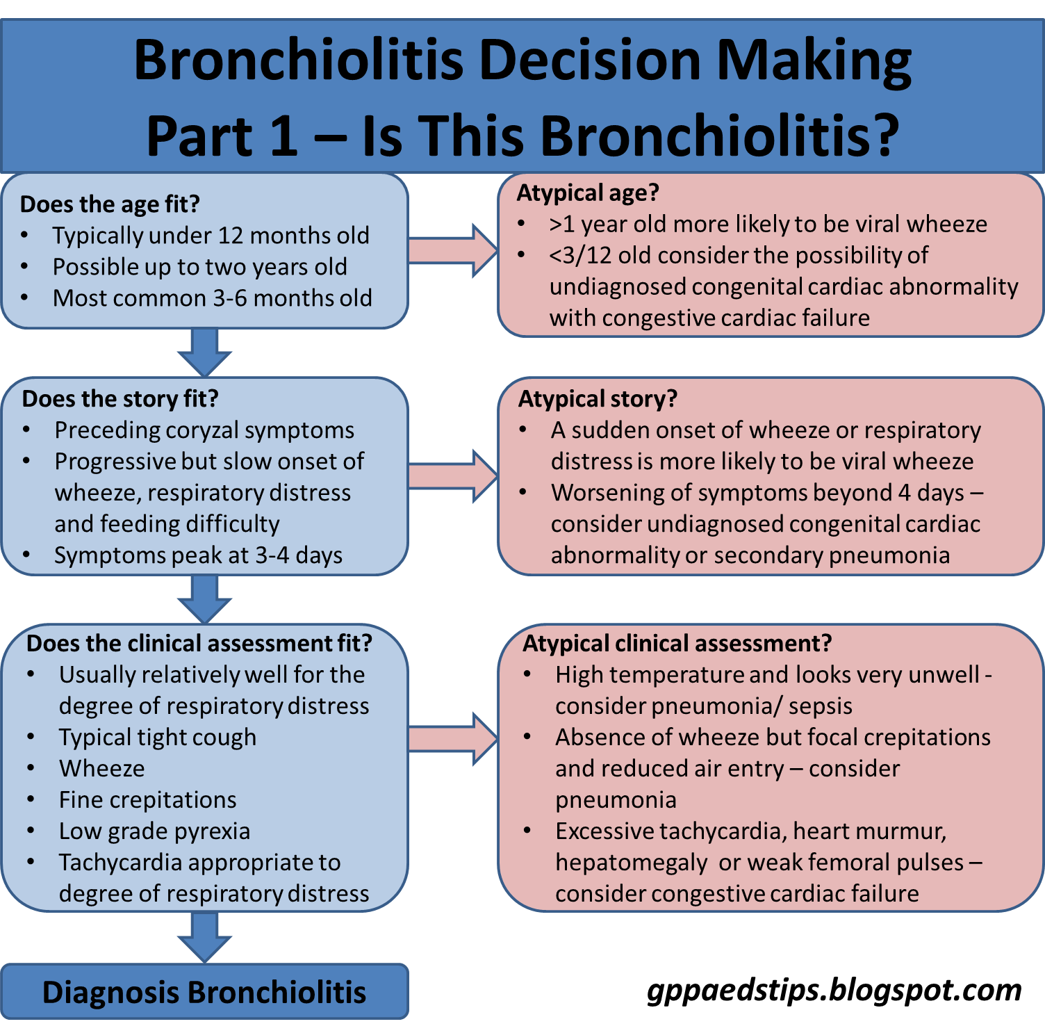
Most children under the age of 1 year presenting with a tight cough, wheeze, respiratory signs and poor feeding have bronchiolitis.  There are other possible explanations for that presentation however and it is important to know about these other possibilities.

Viral induced wheeze, which involves bronchospasm is separate from bronchiolitis.  Clues that it may be viral induced wheeze include the age of the child (most commonly over 1 year) and previous episodes of viral induced wheeze.  The other clue is the onset of the respiratory changes.  Bronchiolitis is a slow accumulation of wetness in the airways and the history is typically of a gradual and progressive worsening of symptoms over days.  Viral induced wheeze, due to the bronchospasm involved, presents with a more sudden onset of wheeze and distress, often going from normal to significantly abnormal over a few hours.

Pneumonia is almost never associated with wheeze in children (2).  Focal crepitations are often heard in a viral lung infection of any kind.  The presence of wheeze strongly suggests that the signs and symptoms are virally induced in some way.  Infants with pneumonia will tend to be significantly unwell.  The simple rule of thumb is this:  If the infant has a wheeze and is well enough to be managed in a pre-hospital setting, they do not have bacterial pneumonia.

Congestive cardiac failure (CCF) due to haemodynamically significant yet undetected congenital cardiac abnormalities is a rare mimic of bronchiolitis but one that is important to be aware of.  The typical cause is a large ventricular septal defect (VSD) causing a significant left to right shunt.  This increased pressure through the lung circulation causes pulmonary odema which manifests as poor feeding, fine crepitations and wheeze.  Thankfully, most significant heart defects are detected before a baby is discharged from postnatal care, but occasionally one slips through and the signs and symptoms are easily mistaken for bronchiolitis.

There are usually clues however.  A murmur is the most obvious clue but this can be difficult to hear at >160bpm.  An excessive tachycardia is a possible sign of CCF.  A significant hepatomegaly (normal babies often have up to a centimetre of palpable liver) is highly suspicious of CCF.  Finally, the progression of symptoms does not fit for bronchiolitis as they continue to get worse after the 3-4 days in which bronchiolitis reaches its peak.

Putting these things together, it is usually possible to be confident in diagnosing bronchiolitis as long as the history and findings are consistent with bronchiolitis and not one of the other pretenders.
If the diagnosis is bronchiolitis, we can move onto our next question:

Question 2: Should this child be managed at home or in hospital?

Most children with bronchiolitis can be managed in the community.  Keeping people away from hospital where it safe to do so has never been more important.  In the UK, the NICE guidelines for bronchiolitis (3) give recommendations for when to refer and when to consider referral.

Referral is always recommended for red flags.  In the NICE guidelines, these are a combination of signs of potential respiratory failure.  Notably, apnoeas are included as a stand-alone red flag.  That means that a child without any chest signs of severe respiratory distress should still be referred if they have had episodes where they appear to stop breathing.  The reason for this is that in such cases, immature respiratory drive may be a factor.  Following an apnoea, a baby can temporarily seem much improved but may go on to have further events and deteriorate suddenly.

Feeding and hydration is probably the least well defined element of the decision making element.  The guidelines ask the clinician to consider a variety of factors, however being able to assess whether the amount of feeding is adequate is next to impossible apart from overt signs of dehydration.  We never know how much a breast fed baby is getting unless the answer is "nothing."  If the baby is bottle fed, applying a percentage to that as being adequate doesn't take into account the fact that many bottle fed babies take much larger volumes as a baseline.  As a result, the most objective measure of adequate feeding has to be signs of hydration or dehydration.  For that reason, I have included clinical dehydration in the list of red flags and beyond that, feeding difficulties remain a matter of clinical judgement when it comes to referral.

Possibly the most controversial element of the decision making is the presence of risk factors.  In the guidance, it is stated "When deciding whether to refer a child with bronchiolitis to secondary care, take account of any known risk factors for more severe bronchiolitis such as... (e.g.) premature birth, particularly under 32 weeks."  The guideline evidence statement lists the basis for each risk factor listed and with the exception of neuromuscular disease, the committee acknowledged that there is no credible published evidence for the other risk factors.  Apart from neuromuscular disease, they are all consensus opinion recommendations.

So what are you supposed to do when you see an 8 month old baby with mild bronchiolitis, no red flags and adequate feeding when you know that they were born at 31 weeks gestation?  Do you send them to secondary care in case because they have a risk factor for severe bronchiolitis or do you keep them well away from hospital because they don't have severe bronchiolitis and you don't want to add a hospital acquired infection to their list of problems?

Balancing risk vs benefit is what it is all about here.  There is a known risk of hospital acquired infection vs an unknown risk of severe bronchiolitis.  There is also no evidence that admitting high risk children with bronchiolitis is any safer than good safety-netting advice.

If the decision is made to manage a child with bronchiolitis at home, the third and final question is:

Question 3: What treatment should the child be given?

There has been a load of research done to try to find an effective treatment for bronchiolitis.  Supportive interventions (oxygen, CPAP etc) in the cases where respiratory support are needed have been shown to be effective.  Each and every other therapy have in turn shown to have no benefit for mild to moderate uncomplicated bronchiolitis.  Therapies proven to be ineffective include β-agonists, ipratopium, hypertonic saline, antibiotics and corticosteroids.  The bottom line is that for a child being managed in the community, no pharmacological treatment should be given.  This recommendation is consistent  across guidelines from the UK, USA and Australia (3,4,5).

That makes this flowchart nice and simple:

Finally, you might be asking yourself if you are an expert decision maker when it comes to a small person who has a cough and wheeze.  Hopefully this post helps you to feel that you are.  Decision making in such children is all about recognition, knowing the red flags and above all, learning that if in doubt, looking at the child will almost always give you your answer.

Edward Snelson
@sailordoctor

References

  1. Roland D, Teo KW, Bandi S, et al COVID-19 is not a driver of clinically significant viral wheeze and asthma Archives of Disease in Childhood Published Online First: 16 October 2020. doi: 10.1136/archdischild-2020-320776
  2. Hirsch A, Monuteaux M, Neuman M, Bachur R, Estimating Risk of Pneumonia in a Prospective Emergency Department Cohort, Paediatrics, Vol 204, p172-176.E1, Jan 01, 2019 doi:10.1016/j.jpeds.2018.08.077
  3. Bronchiolitis in children: diagnosis and management, NICE guideline [NG9] Published date: 01 June 2015
  4. American Academy of Pediatrics Clinical Practice Guideline: The Diagnosis, Management, and Prevention of Bronchiolitis, Pediatrics November 2014, 134 (5) e1474-e1502; doi: 10.1542/peds.2014-2742
  5. The Royal Children's Hospital Melbourne Clinical Practice Guidelines: Bronchiolitis

Tuesday, 8 May 2018

ECG in children - an amuse bouche (and what to do with one)

Food etiquette is a minefield.  I remember the first time I was presented with an amuse bouche.  I had no idea why it was, let alone what I was supposed to do with it.  One minute later, with the tasty morsel in my belly, I realised I had really over-thought the whole thing.  Also, I was still hungry.  Non-acute paediatric ECG is a lot like that.

Some tests are so simple, usually because they are quantitative.  You do a blood sugar and you get... a blood sugar.  Some tests are much more qualitative, such as Chest X-ray.  Show a chest X-ray to half a dozen radiologists and you may be surprised by the range of interpretations.  ECG in children definitely falls into the second category in that it is a test which requires interpretation.  The interpretation of an ECG is fairly standard, but I've never yet met a standard child or a standard clinician.

There are many reasons why an ECG might be done for a child.  I am not talking about during an acute presentation such as a severe tachycardia or other signs suspicious of a cardiac cause for a child to be unwell.  I am talking about ECG in a child who is well, but had a symptom that warranted an ECG.  If you want to know about the kind of critical care ECG interpretation best done wearing a cape and with underpants on the outside of your trousers, you might like to listen to the PEMplaybook.
If your cape is in the cupboard and underwear is wherever you normally keep it, then ECG is a more fickle friend.  When an ECG is normal normal then that's great but it's not the end of the story.  Often it appears abnormal, because paediatric ECGs look different much of the time.  In a BMJ article about ECGs, the authors write, "Chest pain in children is rarely cardiac in origin and is often associated with tenderness in the chest wall. Electrocardiography is not usually helpful in making a diagnosis, although a normal trace can be very reassuring to the family." (1) That is all very well if with a 12 lead ECG you can tell them that all is normal.

So what is the problem with paediatric ECG?  Well actually there are two problems.  The first is the issue of things that look abnormal and are not.
Much of the differences in paediatric ECG are to do with the initial right sided dominance.  The other thing that can be a factor is physics.  The ECG may show up as LVH, RVH, atrial enlargement etc, but this is often because there is just very little in the way of chest wall between the sticker and the myocardium.  You don't get this problem in children with a more substantial chest wall.  Simply put, in a small or skinny child, large waves are usually normal. If something appears big, look at the child and check the axis.  A skinny child and a normal axis means that the "LVH by voltage criteria" is probably a lie.

Knowing these things helps us to be able to say more often, "This is a normal ECG."

Then there is the opposite problem: a 12 lead ECG done when asymptomatic does not rule out significant pathology.  Take this case study as an example:

A 12 year old girl presents having had a collapse while playing tennis.  She had no palpitations or chest pain and simply recalls feeling faint just before she collapsed.  The adult playing tennis with her describes a sudden collapse, while she was walking t pick up a ball.  When they ran over, the young person was unresponsive for only a few seconds before slowly coming around.

Clinical examination is normal.  The only other history of note is in the family history - a sudden unexpected death in infancy of a 9 month old sibling.  

Would you be happy to rule out a cardiac arrhythmia based on a resting, asymptomatic 12 lead ECG?  With that history, I wouldn't recommend it.

So, if an ECG in a child who is currently asymptomatic has a lot of false positives and false negatives, what is it useful for?  The answer to that is that it should mainly be used to answer specific questions.  For example, in the case above, I want to know the corrected QT interval.  A 12 lead ECG will tell me that.  In fact, resting asymptomatic ECGs are mostly useful for checking rhythm and intervals.

Morphology and high voltage account for most of the false positives and normal rhythm can be a false negative.  Both these things are fine, because a 12 lead ECG is simply an amuse bouche.  If you aren't hungry (no red flags), an amuse bouche is simply a tasty mouthful which won't fill you up.  It is debatable whether it is even needed in a child who has had a typical faint with no red flags.

If you are hungry (red flags in the history or examination), don't rely on an amuse bouche, which should just be there to keep you happy until the real food arrives.  What you have for your main course depends on where you work.  Perhaps you have direct access to 24 hr ECG and cardiac echo. Me? I phone a friend to do these things for me.

Edward Snelson
The Gourmand of Child Health
@sailordoctor

Disclaimer - I may not have had all of the ECGs the right way up.  That could explain some of the abnormal morphology.

References
  1. Steve Goodacre, Karen McLeod, Paediatric electrocardiography, BMJ 2002;324:1382
  2. PEM playbook EKG killers


Tuesday, 8 August 2017

Heart Murmurs in Children

Heart murmurs are a reasonably common finding in children.  Excluding the newborn and early infancy assessments, most of the murmurs that we hear are benign, physiological or flow murmurs.  All of those terms basically mean the same thing – the anatomy is normal, the child is healthy and yet there is an extra noise heard on auscultation of the heart.  The reason that this usually occurs is that children’s physiology responds vigorously to illness and stress.  The heart pumps faster and harder, often making a structurally normal heart into a noisy heart.

In short, heart murmurs are almost always nothing to worry about.  Almost.

The trouble is that everything in paediatrics has an evil twin.  So what are the possible more significant  pathologies and how can these be recognised amongst the far more common benign diagnoses?  When children present with an acute illness, a cardiac cause for their symptoms is not usually top of our list of differential diagnoses because respiratory and musculoskeletal cases are far more common reasons for chest pain or dyspnoea.  This coupled with the way that cardiac problems present (with vague symptoms easily attributable to more common illnesses) make these rare clinical scenarios into the stuff of our worst fears.


Let’s look at a few made up cases to illustrate how to approach the “Oooh-I’ve-heard-a-murmur-what-now?” scenario.

Case 1

A 3 year old child presents with a febrile illness.  Let’s say that they have an otitis media.  They have a heart rate at the top of the reference range for their age and you hear a murmur.  The murmur is soft, easily heard (but not loud), systolic and heard best at the left sternal edge.  There is no radiation.

What now?

In primary care, we are all about the focussed history and examination.  We have to be in order to make time for our other patients.  That ergonomic approach works well, but when we find something that we weren’t expecting, we need to go back and get more information.  In this case, we want to know if the child is known to have a heart murmur.  Have they had an echo done in the past?  We also need to feel the precordium for heaves and thrills and be happy that the pulses have a normal character and volume.  Make sure that the femoral pulses are palpable, with no brachio-femoral delay.  Check for hepatomegaly.  Most importantly, make sure that the child does not have increased work of breathing and does not seem unexpectedly unwell.  Ideally, we need to get a blood pressure checked.

What next?

We need to refer a child with a murmur acutely (usually to general paediatrics) if

  • they are more unwell than expected
  • they have respiratory symptoms which are otherwise unexplained
  • there are concerning clinical examination finding (e.g. hepatomegaly)

A well child with none of the above ‘red flags’ probably needs an outpatient echo if

  • The murmur is loud
  • The murmur is diastolic
  • The murmur radiates outside of the precordial area

If the child is well, there are no red flags and the murmur sounds benign (as in the original description for this case) then common practice is to follow the child up when they are well again.  The presumption is that this is a physiological murmur which has been heard because the illness is causing increased cardiac output and therefore turbulent blood flow.

It is likely that when they are seen, the murmur will no longer be heard.  In these circumstances: case closed.  If the murmur persists, they can be referred then (either for an echo or to paediatrics if they don’t have direct access to this), assuming that the child remains red-flag-free.

Case 2

A two month old baby presents with a runny nose, cough and a low grade temperature.  The parent has noticed that their baby has started to have slightly fast breathing and has not been feeding as well as they normally do.  On examination, the baby has a slight wheeze, mild recession and basically looks like the other babies with bronchiolitis that you have seen that month.  All except for one thing: they have a soft systolic murmur that you can just about hear over the wheeze.

What now?

In short, refer to the acute paediatric medical team.  This child might have bronchiolitis and a flow murmur, but there is every chance that they are just pretending to have bronchiolitis.  Babies who have a ventricular septal defect (VSD) might not be picked up on screening (newborn baby checks etc.) and may have no overt symptoms, until they get their first cold.  Then, shortly after becoming snotty, the illness tips them into heart failure.  What does heart failure look like in a baby?  Well, they have increased work of breathing, a bit of a wheeze and struggle to feed.  Does that sound like bronchiolitis or does that sound like bronchiolitis?

Other clues that give these mimics away are:

  • Excessive tachycardia
  • Hepatomegaly
  • More pale or mottled
  • The course of the illness is different – they continue to get worse, while bronchiolitis symptoms peak at day 3-4 of the illness.

Case 3

A fourteen year old presents a few days into a flu-like illness.  They have been seen by a couple of doctors in the past few days, and have been told that they have a viral illness.  On both occasions they were advised about symptomatic treatment.  The parents are concerned that the young person is not getting better.  On examination, there is a barely audible systolic murmur.

What now?

Don’t presume this to be a flow murmur.  Firstly, in the context of viral illness, flow murmurs are most commonly heard in younger children. Also, the the murmur was not heard on previous visits.   One possible diagnosis here is viral myocarditis.  Other possibilities include bacterial endocarditis.

What next?

Refer this child acutely to the paediatric medical team.


One general rule which works quite well for symptoms in children (in the context of heart problems) is this: An isolated sign or symptom is rarely indicative of significant pathology.  Two signs or symptoms is always worth taking very seriously.  Take chest pain in children.  I don’t expect chest pain to be due to a cardiac cause in children (because it almost never is).  I know that the vast majority of children who have a syncopal episode will be having straightforward vasovagal events.  However, chest pain in combination with syncope, dyspnoea or palpitations is presumed by me to be pathological until said pathology is excluded.


So, as with many presentations in children, everything is normal except when it isn’t.

Edward Snelson
Postcordiologist
@sailordoctor


Many thanks to Dr. Carrie Mackenzie (Consultant paediatrician) who helped with the original version of this piece which is on the excellent RCEM learning site.


Thursday, 20 July 2017

The Everest-Lifeboat Test (Easter Egg - Investigating apparent faints in children)

Paediatric guidelines always have to take into account one of the most important maxims of Child Health - the test or treatment must always be in the best interest of the child.  This means a particular discernment about the value of a test or treatment.  Guidelines rarely discriminate between which treatments and tests are essential and which are simply recommended.  When writing a guideline, it is relatively easy to put in a recommendation, often giving the impression that it is a must-do when this is simply not the case.


One of the things that I am most often asked (whether at work or giving and educational session) is "Do I have to do X?  The guideline says to but..."  So how does one discriminate between the must-do's and the would-be-good-if we-could recommendations?  That's easy - apply the Everest-Lifeboat Test.

The Everest-Lifeboat Test is simply a two part thought exercise.  The first part is to ask the following question:  If the person involved was at an advanced stage of climbing Everest, should they turn back to get this test or treatment or could they reasonably continue to the summit?  This part of the test asks if we are doing something just because we can rather than because we have to.  We are purely focusing on the immediate need at this point.

The second part is the lifeboat question:  Imagine that this patient was in a lifeboat, drifting across the ocean.  When they are rescued several weeks later, would they most likely be fine despite their current clinical situation?  This part of the Everest-Lifeboat Test forces us to look forward and consider the medium and long term consequences of action versus inaction.

Let's try this on a fictional patient.

A 10 year old child is brought to you having had an unexplained collapse.  The history given to you is vague and there is no first hand account of the episode as it happened in front of classmates at school.  However, in your further history taking, you find that this child has been having headaches that are worse in the morning.  Also, teachers have noticed that there has been a deterioration in performance at school over the past four weeks.  The child is slightly ataxic and has nystagmus on examination.

Concerned, you discuss the need for further investigations.  In response, the parents say that they are happy to get the tests done but could it please wait a few weeks?  They were just coming to get a medical opinion before they catch a plane to go away for a couple of weeks for their holiday.  They were assuming that this was just a fainting episode and really only came to get their child checked because grandma told them to.

So, what do you think?  This seems like a clear fail of the Everest-Lifeboat Test to me.  I wouldn't be happy to put investigations on hold, despite the obvious inconvenience to the family.

How about this scenario?:

A 12 year old girl comes to be assessed.  She was in school and had been stood in the heat when she began to feel sweaty and nauseous.  Her vision went black and she slumped to the floor.  She was reported to look pale and floppy.  She was unresponsive for a few seconds and then came around slowly over a few minutes.  A few hours later, she feels fine.  There is no history of unexplained deaths in her family.  When you examine her, all is normal.

You check a relevant guideline and see that it recommends that you perform a 12 lead ECG. She flatly refuses to have this test done and will not be persuaded.  What should you do?

Applying the Everest-Lifeboat test would go like this:

Would you advise abandoning the final attempt on the summit?  Well, she had an obvious precipitant and prodrome for her apparent faint.  We can advise how to avoid precipitants and what to do if a prodrome is recognised.  The event appears to be a classical vasovagal syncope without red flags in the history or examination.  So, forcing the issue seems to be unnecessary.

Would a few weeks in a lifeboat be an issue?  This brings us back to the guidelines that recommend investigation.  What are they trying to protect us from?  Much of the practice of ECG screening comes from adult medicine, where pathology is much more likely.  In paediatrics, there are a few arrhythmias that we need to worry about, but a standard 12 lead ECG is not the perfect screening test that we might hope it is.  The sensitivity and specificity of 12 lead ECGs in children is poor (1).  Ask yourself why the guidelines don't say, "Don't bother with history and examination.  The ECG is the crucial bit of information."

A quick look at the guidelines gives some useful insights to help us with the Everest-Lifeboat Test.  The NICE guideline "Transient loss of consciousness (‘blackouts’) management in adults and young people" (2) actually only relates to the over 16 year-olds.  This in itself acknowledges that an adult approach cannot be extrapolated to the child who has had a collapse.

Then there is the European Society of Cardiology's guideline for the diagnosis and management of syncope (version 2009). (3) It does take the view that children and adults can be investigated similarly and recommends ECG for all children who have had a faint.  However, this recommendation seems to be based on the assumption that an ECG is clearly useful additional information and fails to consider the possibility that a thorough history and examination gets you to a point where and ECG would not add value.

The American College of Cardiology/American Heart Association guideline (4) does seem to consider this possibility.  There is a strong emphasis on the value of a good history and examination.  Regarding ECG, it points out that ECG is a simple and available test that might identify a tendency to arrhythmia.  However it also states: "Despite the benefit of identifying a likely cause or potential clue about the cause of syncope from the ECG, prospective studies did not conclude that ECG findings significantly affected subsequent management.  The prognostic value of an abnormal ECG in patients with syncope has been questioned as well."

So there it is.  An honest declaration that, while the experts would recommend that we all do a test, it is unclear what the value of the test is.

I know that it might seem as though I just want to avoid doing work here, but there are genuine risks with tests.  The first risk is that they stop us from thinking.  If the sensitivity and specificity of history and examination is excellent, while that of ECG is poor, why introduce a deceptive piece of information?  The second risk is that of getting information that I don't want.  If I do an ECG on a child, it is almost always to look at the rate, rhythm and QT interval.  While those things are usually fine, the diagnostic report usually sports a bit of LVH and right atrial enlargement.  Of course the child has neither of these things, but the machine is just trying to make sense of the voltage it has been given.  If I were to take these things seriously, I might cause unnecessary anxiety for the child and parents.

So, what does my patient really need?  I need to take a good history and establish that the episode that sounds like a faint truly sounds like a faint.  This means asking about the three P's of vasovagal syncope.
If it sounds like a classic faint, I still need to make sure that I consider my red flags.
If the history given is of a straightforward faint, without red flags, I think that allowing the child to refuse the ECG passes the Everest-Lifeboat Test.

When we are forced out of our normal process, it is a good time to evaluate our routine practice.  If a deviation from the norm passes the Everest-Lifeboat Test, I would question the norm.  You may just have discovered that you are doing a test or a treatment that you don't believe in.  Here is a little list of things that have passed the Everest-Lifeboat Test for me at various times in the past (i.e. I was going to treat, something got in the way of that and I went with the the no treatment option):
That's a short list of times when the option of doing nothing became the right thing despite what was routine practice.  In the case of umbilical granuloma, I am pleased to say that doing nothing is now becoming the norm.

I hope that you find the Everest-Lifeboat Test useful at some point.  As to whether every child who has had a faint needs an ECG, versus it is good to get one or it is simply not needed unless there is a specific reason - this is a debate that is lacking input from the good people of the primary care and emergency medicine communities.  My solution?  Stick a cardiologist, a paediatrician, an emergency medicine doctor and a general practitioner in a lifeboat and leave them there till they've sorted it out.  I'm fairly sure they'd be fine...

Edward Snelson
Precordiologist
@sailordoctor

Disclaimer: The Everest-Lifeboat Test was originally described in 1055 by Egbert the Uncertain, a monk who died at the Battle of Hastings before writing down his idea.  I therefore take full credit for inventing the test myself.


  1. Kapoor WN, Evaluation and outcome of patients with syncope, Medicine, 1990 May;69(3):160-75.
  2. NICE guideline "Transient loss of consciousness (‘blackouts’) management in adults and young people"
  3. Diagnosis and management of syncope, European Society of Cardiology, European Heart Journal (2009) 30, 2631–2671
  4. Guideline for the Evaluation andManagement of Patients With Syncope,  A Report of the American College of Cardiology/American Heart Association Task Force on Clinical Practice Guidelines, and the Heart Rhythm Society